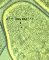
F. leucas 500-550 μm Intact cells Japan, 199? |

F. leucas 928 x140 μm trich. 9 x 1.5 μm ingesting food Ichikawa Chiba, 2001 |

F. leucas 540-800 μm Koaze river Kawagoe Saitama, 2002 |

F. leucas ? Intact cells Japan, 199? |

F. leucas stock, Nnm-1 280-312 μm trich. 8 x 1 μm Matsumoto Nagano, 2001 |
| DV images | ||||

Nnm-1 348 x μm 4-9 Mic. Matsumoto Nagano, 2001 |

Cos-2 353 x μm 3-7 Mic. Ichikawa Chiba, 2001 |

Cos-1 391 x μm 3-4 Mic. Ichikawa Chiba, 2001 |

Gmt-6 369 x μm 2-4 Mic. Tatebayashi Gunmma, 2001 |

Hsf-4 μm Sanda Hyogo, 2001 |

SIR-24 μm Iruma Saitama, |

Ckr-1 μm 0 Mic. ? |
|||
Misc.

F. leucas ? F. marina ? Intact cells Japan, 199? |

F. leucas ? cell division Japan, 199? |

F. leucas ? cyst formation? Japan, 199? |

F. leucas ? excystment ? Japan, 199? |

Frontonia sp. F. marina ? Intact cells Japan, 199? |

Frontonia sp. F. marina ? Mac. and Mic. Japan, 199? |

Frontonia sp. Mac. Japan, 199? |

Frontonia sp. Mac. & Mic. Japan, 2002 |
